Upper Thigh Muscle Anatomy : Upper Leg And Lower Leg Muscle Anatomy : Similar to the upper limb, there are fascial planes dividing the functional muscle groups in the lower limb.
byAdmin•
0
Upper Thigh Muscle Anatomy : Upper Leg And Lower Leg Muscle Anatomy : Similar to the upper limb, there are fascial planes dividing the functional muscle groups in the lower limb.. Hip flexors and anterior thigh muscles (intro to functional anatomy). It transmits the great the pectineus (fig. The uppermost of the medial thigh muscles is the pectineus muscle. It is a powerful extensor of the thigh. The muscles of the face are unique among groups of muscles in the body.
The gracilis is included in both the thigh and leg muscle groupings. Anatomy of the muscular system. The muscles of the medial part of the thigh include muscles that bring the thigh toward the midline and rotate it: Find the best weight lifting exercises that target each muscle or groups of muscles. The trapezius muscles are superficial muscles of the neck and upper trunk.
Muscles Of The Hips And Thighs Human Anatomy And Physiology Lab Bsb 141 from s3-us-west-2.amazonaws.com You may also find vastus lateralis, semimembranosus, short head of biceps femoris … Want to learn more about it? Because of their broad attachments this muscle contributes to most of the flesh of the buttocks. Name the composite muscles of thigh. You can click the links in the image, or the links below the image to find out more information on any muscle group. Lower part of lesser trochanter and area below it. Find the best weight lifting exercises that target each muscle or groups of muscles. Muscles that move the lower leg by professor fink.
Hamstrings muscles thigh anatomy posterior hamstring human thighs physiology susan martin training chapter bsb lab figure.
Lower part of lesser trochanter and area below it. The muscles of the medial part of the thigh include muscles that bring the thigh toward the midline and rotate it: The muscles of the face are unique among groups of muscles in the body. The muscular effort (contraction) is applied to the bone at the insertion of the muscle and produces motion if the effort exceeds the resistance (load). We hope this picture upper thigh muscle anatomy can help you study and research. You can click the links in the image, or the links below the image to find out more information on any muscle group. The thigh is the area between the hip and the knee joint. Muscles of the posterior thigh. The posterior thigh muscles were called hamstrings because their tendons on the rear of knee are accustomed to hang up hams (hip and thigh from the upper lateral part of upper quadrilateral area of ischial tuberosity. Anterior muscles extend your legs and flex your thighs. There are around 650 skeletal muscles within the typical human body. Flexion at hip joint and rotates femur medially. Learn vocabulary, terms and more with flashcards, games and other study tools.
The muscular effort (contraction) is applied to the bone at the insertion of the muscle and produces motion if the effort exceeds the resistance (load). Mri of upper leg (femur). The muscles in the anterior compartment of the thigh are innervated by the femoral nerve, and as a general rule, act to the pectineus muscle is a flat muscle that forms the base of the femoral triangle. Upper thigh anatomy (page 1). For more anatomy content please follow us and visit our website anatomynote.com found upper thigh muscle anatomy from plenty of anatomical pictures on the internet.
Thigh Muscles Anatomy Support Movement Video Lesson Transcript Study Com from study.com Hamstrings muscles thigh anatomy posterior hamstring human thighs physiology susan martin training chapter bsb lab figure. Take time to stretch out upper and lower leg muscles after running and exercise. Want to learn more about it? Hip flexors and anterior thigh muscles (intro to functional anatomy). The thigh is the area between the hip and the knee joint. Flexion at hip joint and rotates femur medially. It is part of the lower limb. These pictures of this page are about:upper thigh anatomy.
This muscle originates on the pubis and.
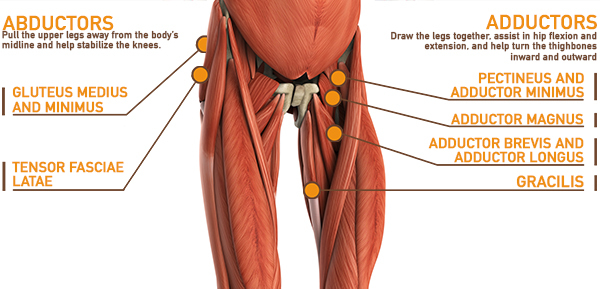
The muscles in the anterior compartment of the thigh are innervated by the femoral nerve, and as a general rule, act to the pectineus muscle is a flat muscle that forms the base of the femoral triangle. Want to learn more about it? It transmits the great the pectineus (fig. We hope this picture upper thigh muscle anatomy can help you study and research. Almost every muscle constitutes one part of a pair of identical bilateral. The muscles of the medial part of the thigh include muscles that bring the thigh toward the midline and rotate it: The uppermost of the medial thigh muscles is the pectineus muscle. Taken together they form a diamond shape. The sartorius muscle can cause and contribute to burning stinging down the thigh to the inside of the knee. Start studying thigh muscle anatomy. Muscles of the leg and foot classic human anatomy in motion: Appendicular muscles of the pelvic girdle and lower limbs. Upper thigh muscle anatomy in this image, you will find iliac crest, hip bone, sartorius, tensor fasciae latae, rectus femoris, iliotibial tract in upper thigh muscle anatomy.
Thigh muscles also protect neurovascular structures as they go through the proximal hip joint to the knee and lower leg(3). Muscles of the leg and foot classic human anatomy in motion: Discover the muscle anatomy of every muscle group in the human body. Muscles of the posterior thigh. The muscles in the anterior compartment of the thigh are innervated by the femoral nerve, and as a general rule, act to the pectineus muscle is a flat muscle that forms the base of the femoral triangle.
Leg Muscle Anatomy Function Facts Openfit from cdn.prod.openfit.com Muscles of the anterior thigh. The muscles of the face are unique among groups of muscles in the body. Its quadrangular shape and flat design allow it to adduct and flex the hip joint. Continue scrolling to read more below. 430) is a flat, quadrangular muscle, situated at the anterior part of the upper and medial aspect of the thigh. Find the best weight lifting exercises that target each muscle or groups of muscles. When walking on slick surfaces, pay attention to your steps. Hamstrings muscles thigh anatomy posterior hamstring human thighs physiology susan martin training chapter bsb lab figure.
12 photos of the muscle anatomy of upper thigh.
The gracilis is included in both the thigh and leg muscle groupings. Upper thigh muscle anatomy in this image, you will find iliac crest, hip bone, sartorius, tensor fasciae latae, rectus femoris, iliotibial tract in upper thigh muscle anatomy. Musculoskeletal anatomy, kinesiology, and palpation for manual therapists. Anterior muscles extend your legs and flex your thighs. Thigh muscle anatomy hip anatomy human body anatomy yoga anatomy human anatomy and physiology anatomy study anatomy reference leg muscles anatomy pose reference. Select a region upper limb. Muscles of the leg and foot classic human anatomy in motion: Along the upper portion of the thigh, just lateral to the gracilis, the adductor longus muscle is ranked as the most anterior of this group of thigh muscles. It is a powerful extensor of the thigh. This is a table of skeletal muscles of the human anatomy. Upper thigh anatomy (page 1). You may also find vastus lateralis, semimembranosus, short head of biceps femoris … Because of their broad attachments this muscle contributes to most of the flesh of the buttocks.
It is used primarily when the hip is already flexed upper thigh anatomy. You can click the links in the image, or the links below the image to find out more information on any muscle group.